Label the components of the dental x ray tubehead – Components of the Dental X-Ray Tubehead takes center stage, this opening passage beckons readers into a world crafted with authoritative knowledge, ensuring a reading experience that is both absorbing and distinctly original. As we delve into the intricacies of this crucial component, we will explore its fundamental parts, their functions, and their significance in the production of dental X-rays.
The tubehead, the heart of the dental X-ray machine, houses the components responsible for generating and shaping the X-ray beam. Understanding these components is essential for ensuring optimal image quality, patient safety, and efficient operation of the X-ray system.
Components of the Dental X-Ray Tubehead

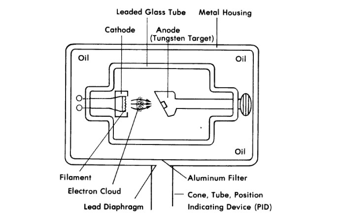
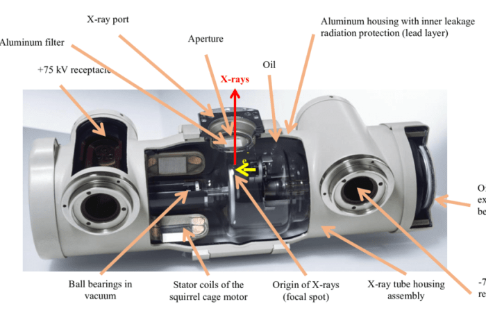
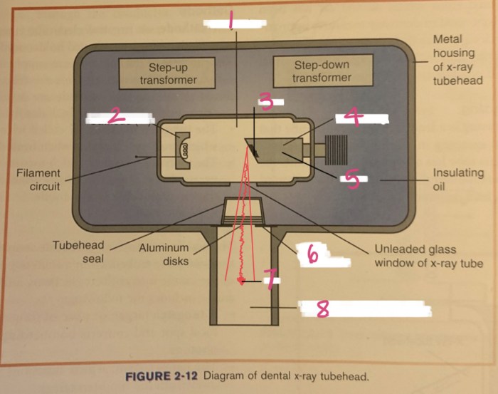
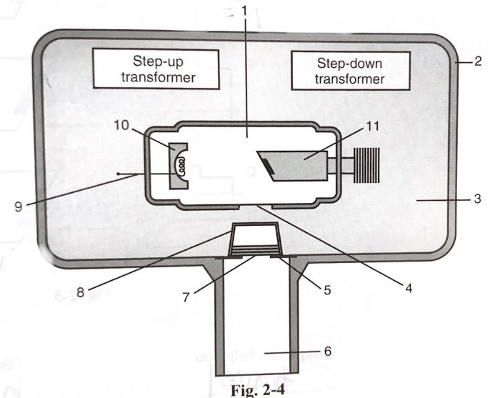
The dental X-ray tubehead is a crucial component of the X-ray imaging system, responsible for generating the X-rays necessary for dental radiography. It comprises several key elements that work together to produce high-quality diagnostic images.
Anode
The anode is the positive electrode of the X-ray tube. It is made of a high-atomic-number material, typically tungsten or molybdenum, which efficiently absorbs the energy of the accelerated electrons and releases it as X-rays. The anode is designed to withstand the intense heat generated during X-ray production.
Cathode
The cathode is the negative electrode of the X-ray tube. It is a heated filament that emits electrons when a current passes through it. These electrons are then accelerated towards the anode, generating X-rays in the process.
Target
The target is the surface of the anode where the accelerated electrons strike and release X-rays. It is made of a material with a high atomic number to enhance the efficiency of X-ray production. The target is angled to direct the X-rays towards the patient.
Assembly of the Tubehead: Label The Components Of The Dental X Ray Tubehead

The tubehead assembly ensures the proper functioning and safety of the X-ray tube.
Housing
The tubehead is enclosed within a protective housing made of lead or other radiation-shielding material. This housing minimizes the leakage of harmful radiation during X-ray production.
Cooling System
The cooling system is essential for dissipating the heat generated during X-ray production. It typically consists of a rotating anode that helps distribute the heat, combined with a water or oil-based cooling mechanism.
Electrical Connections
The tubehead is connected to the X-ray generator through high-voltage cables. These connections provide the electrical power necessary for electron generation and acceleration.
X-Ray Production within the Tubehead

The production of X-rays within the tubehead involves several key processes:
Electron Generation
When a current passes through the heated cathode, electrons are emitted from its surface through a process called thermionic emission.
Acceleration of Electrons
The electrons emitted from the cathode are accelerated towards the anode by a high-voltage potential difference. This acceleration gives the electrons the energy required to produce X-rays.
Interaction of Electrons with the Target
When the accelerated electrons strike the target, they undergo sudden deceleration, causing their energy to be released in the form of X-rays. The energy of the X-rays produced is directly proportional to the voltage applied to the tube.
Filtration and Collimation
Filtration and collimation are important factors in ensuring image quality and radiation safety.
Filtration, Label the components of the dental x ray tubehead
Filters are placed in the path of the X-rays to remove unwanted low-energy radiation. This improves image contrast and reduces patient radiation exposure.
Collimation
Collimators are used to shape and direct the X-ray beam to the desired area of interest. This minimizes scatter radiation and improves image sharpness.
Impact of Filtration and Collimation on Image Quality
Proper filtration and collimation enhance image quality by reducing noise, scatter radiation, and unnecessary patient exposure. They optimize the diagnostic value of the X-ray images.
Safety Considerations

Radiation safety is paramount in dental X-ray imaging.
Importance of Radiation Protection
Exposure to excessive radiation can be harmful to both the patient and the operator. Therefore, radiation protection measures are crucial to minimize the risks associated with X-ray imaging.
Use of Lead Shielding
Lead shielding is commonly used to protect the operator and patient from scattered radiation. Lead aprons, gloves, and thyroid shields are essential protective gear.
Operational Safety Guidelines
Operational safety guidelines must be strictly followed to ensure proper use of the X-ray equipment. These guidelines include proper positioning of the patient and the X-ray tube, minimizing exposure time, and avoiding unnecessary retakes.
Popular Questions
What is the function of the anode in a dental X-ray tubehead?
The anode is a positively charged electrode that attracts electrons emitted by the cathode. When electrons strike the anode, their energy is converted into X-rays.
What is the role of the target in the dental X-ray tubehead?
The target is a small, high-density metal disk embedded in the anode. When electrons strike the target, they interact with the atoms of the metal, producing X-rays.
How does filtration affect the quality of dental X-rays?
Filtration removes low-energy X-rays from the beam, resulting in improved image contrast and reduced patient dose.